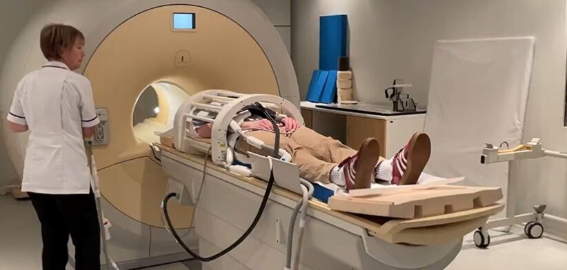
A new medical scan can monitor lung function in real-time allowing for better treatment of pulmonary conditions.
Researchers at Newcastle University, in the United Kingdom, said the new method could also show experts how transplanted lungs were functioning.
Project lead Pete Thelwall said with the new scan method researchers could see how air moved in and out of the lungs as people took a breath.
Professor Thelwall said it was used in patients with asthma, chronic obstructive pulmonary disease (COPD), and patients who had received a lung transplant.
He said the scan used a special gas that could be seen on an MRI scanner.
“The gas can be safely breathed in and out by patients, and then scans taken to look at where in the lungs the gas has reached.
“Our scans show where there is patchy ventilation in patients with lung disease, and show us which parts of the lung improve with treatment.
“For example, when we scan a patient as they use their asthma medication, we can see how much of their lungs and which parts of their lung are better able to move air in and out with each breath.”
Professor Thelwall said using the new scanning method, the team were able to reveal the parts of the lung that air didn’t reach properly during breathing.
He said by measuring how much of the lung was well-ventilated and how much was poorly ventilated, experts could make an assessment of the effects of a patient’s respiratory disease, and they could locate and visualise the lung regions with ventilation defects.
Read the full study: Assessing Lung Ventilation and Bronchodilator Response in Asthma and Chronic Obstructive Pulmonary Disease with 19F MRI.


