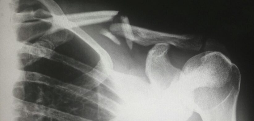
A world-first method to interpret X-rays has been developed by Australian scientists.
A team at the CSIRO have trained artificial intelligence (AI) to write more accurate chest X-ray reports by giving it the same information doctors use in real life.
Study lead author Aaron Nicolson said the team used more than 46,000 real-world patient cases from a leading US hospital dataset to teach a powerful multimodal language model to generate detailed radiology reports.
Dr Nicolson said the results showed 17 percent better diagnostic insights and stronger alignment with expert radiologist reporting.
He said with hospitals worldwide struggling to keep pace with demand amid chronic radiologist shortages, the research could pave the way for faster, safer, and more reliable X-ray reporting in clinical settings.
“Until now, AI tools tasked with interpreting chest X-rays relied solely on the images themselves and the doctor’s referral, without being equipped to read the vital clues hidden in patients’ medical records.”
Dr Nicholson said the CISRO work flipped that approach, by combining imaging with emergency department data like vital signs, medication history and clinical notes to vastly improve diagnostic performance.
“The AI is functioning as a diagnostic detective and we’re equipping it with more evidence.
“When you combine what’s in the X-ray with what’s happening at the bedside, the AI gets more accurate, and much more useful.”
Read the full study: CXRMate-ED: The Impact of Auxiliary Patient Data on Automated Chest X-Ray Report Generation and How to Incorporate It.